These nerves travel outside of the spinal canal to the upper extremities (arms, hands and fingers), to the muscles of the trunk, to the upper and lower extremities (arms, hands, fingers, legs, feet and toes) and to the organs of the body.Īny interruption of spinal cord function by disease or injury at a particular level may result in a loss of sensation and motor function below that level. The PNS is a complex system of nerves that branch off from the spinal nerve roots. One pair of coccygeal (Co1) nerves meets in the area of the tailbone.īy way of the peripheral nervous system (PNS), nerve impulses travel to and from the brain through the spinal cord to a specific location in the body. The first nerve root exits between S1 and S2. The first of these nerve roots exits between L1 and L2.
/3d-illustration-of-spinal-cord--thoracic-vertebrae--a-part-of-human-skeleton-anatomy-698558388-21ae820feeea4a7aa9631c52747faa43.jpg)
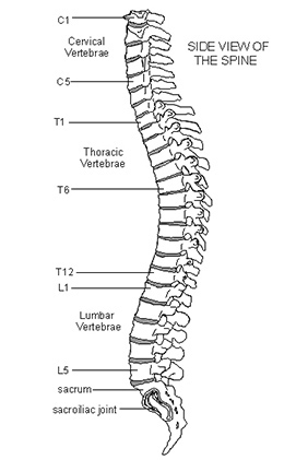
The first nerve root exits between the T1 and T2 vertebrae. The eighth nerve root exits between the C7 and T1 vertebra. The second cervical root exits between the C1-C2 segment and the remaining roots exit just below the correspondingly numbered vertebra. The first cervical root exits above the C1 vertebra. One member of the pair exits on the right side and the other exits on the left. Eight pairs of cervical nerves exit the cervical cord at each vertebral level. There are 31 pairs of spinal nerves and roots. Ligaments attached to the vertebrae also serve as supportive structures. These discs act as shock absorbers for the spinal bones. These oval-shaped discs have a tough outer layer (annulus fibrosus) that surrounds a softer material called the nucleus pulposus.

Five vertebrae are fused together to form the sacrum (part of the pelvis), and four small vertebrae are fused together to form the coccyx (tailbone). The spinal cord lies inside the spinal column, which is made up of 33 bones called vertebrae. A fibrous band called the filum terminale begins at the tip of the conus medullaris and extends to the pelvis.Īt the bottom of the spinal cord (conus medullaris) is the cauda equina, a collection of nerves that derives its name from the Latin translation of "horse's tail" (early anatomists thought the collection of nerves resembled a horse's tail).Ĭerebrospinal fluid (CSF) surrounds the spinal cord, which is also shielded by three protective layers called the meninges (dura, arachnoid and pia mater). The cervical (neck) and lumbar (lower back) segments house the spinal cord's two areas of enlargement. The spinal cord is about 18 inches (45 centimeters) in length and is relatively cylindrical in shape. The spinal cord begins at the bottom of the brain stem (at the area called the medulla oblongata) and ends in the lower back, as it tapers to form a cone called the conus medullaris.Īnatomically, the spinal cord runs from the top of the highest neck bone (the C1 vertebra) to approximately the level of the L1 vertebra, which is the highest bone of the lower back and is found just below the rib cage.

The spinal cord is an extension of the central nervous system (CNS), which consists of the brain and spinal cord.


 0 kommentar(er)
